Scientific Research Division

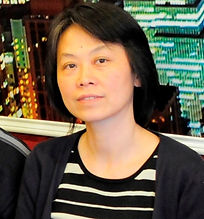
Su-Yu Chiang (江素玉), Ph. D.
Associate Research Scientist
Life Science Group
Office: Room S315, R&D Building
E-Mail: schiang@nsrrc.org.tw,
Tel: +886-35780281-7315
Education
1987-1991 Ph.D. in Physical Chemistry, National Tsing Hua University (Taiwan)
Employment
1994- Associate scientist, SRRC
1993-1994 Postdocral fellow, Department of Chemistry, State University of New York at Stony Brook
1991-1992 Postdoctral fellow, Department of Chemistry, University of California, Berkeley
Research Interest
We are interested in developing high-resolution microscopic techniques for revealing detailed structures of biological samples. We have implemented a structured illumination (SI) based fluorescence imaging system to provide doubled resolution than the diffraction limit. Now, we are working on the developments of coherent SI imaging for native samples without labelling fluorophores and correlated SI-fluorescence microscopy (SIFM) with soft x-ray tomography at Taiwan photon source (TPS) for bio-medical research. Fluorescence imaging that can provide functional information of biological samples is an important complementary method of contrast-based x-ray imaging methods. The use of structured illumination microscopy (SIM) facilitates the system integration to provide improved resolution. With these developments, we will collaborate with biologists to study important fine structures of biological samples to explore new phenomena. Figure shows a system photo and fluorescence images of microtubules taken with wide-field and SIM to show resolution improvement.

Selected Publication
1. Optical imaging system using structured illumination. S.-Y. Chiang*, B.-J. Chang, L.-J. Chou, and J.-Y. Yuh, Japan Patent No. 5524308 (2014).
2. Three-beam interference with circular polarization for structured illumination microscopy. H.-C. Huang, B.-J. Chang, L.-J. Chou, and S.-Y. Chiang*, Optics Express, 21(20), 23963-23977 (2013).
3. Cellular uptake and phototoxicity of surface-modified fluorescent nanodiamonds. M.-F. Weng, B.-J. Chang, S.-Y. Chiang*, N.-S. Wang and H. Niu, Diamond Relat. Mater. 22, 96-104 (2012).
4. Subdiffraction scattered light imaging of gold nanoparticles using structured illumination. B.-J. Chang, S. H. Lin, L.-J. Chou, and S.-Y. Chiang*, Optics Letters 36, 4773 (2011).
5. Probing the binding kinetics of proinflammatory cytokine-antibody interactions using dual color fluorescence cross correlation spectroscopy. C.-Y. Wu, C.-K. Huang, C.-Y. Chung, I-P. Huang, Y. Hwu, C.-S. Yang, L.-W. Lo*, S.-Y. Chiang*, Analyst 136, 2111 (2011).
6. Isotropic image in structured illumination microscopy patterned with a spatial light modulator. B.-J. Chang, L.-J. Chou, Y.-C. Chang and S.-Y. Chiang*, Optics Express. 17, 14710 (2009).
7. Dissociation of energy-selected c-C2H4S+ in a region 10.6-11.8 eV: threshold photoelectronphotoion coincidence experiments and quantum-chemical calculations. Y.-S. Fang, I-F. Lin, Y-C. Lee, and S.-Y. Chiang*, J. Chem. Phys. 123, 054312 (2005).
8. Experiments and quantum-chemical calculations on Rydberg states of H2CS in the region 5.636-9.537 eV. S.-Y. Chiang* and I.-F. Lin, J. Chem. Phys. 122, 94301 (2005).
9. Experimental and quantum-chemical studies on photoionization and dissociative photoionization of CH2Br2. S.-Y. Chiang*, Y.-S. Fang, M. Bahou, and Y.-P. Lee, J. Chem. Phys. 120, 3270 (2004).